Fig. 7
- ID
- ZDB-FIG-111020-16
- Publication
- de Borsetti et al., 2011 - Light and melatonin schedule neuronal differentiation in the habenular nuclei
- Other Figures
- All Figure Page
- Back to All Figure Page
|
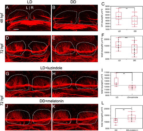
Neuropil formation in the habenular nuclei is promoted by melatonin. (A–C) At 48 hpf, the amount of dense neuropil in the left habenular nucleus is reduced by 28.5% in DD conditions, relative to LD controls. (D–F) At 72 hpf, total neuropil in both habenular nuclei is reduced by 21% in DD conditions, relative to LD controls. (G–I) Treatment with luzindole under LD conditions causes a reduction in neuropil similar to DD conditions (2 examples are shown). (J–L) Conversely, treatment with melatonin under DD conditions rescues neuropil density to be similar to LD (2 examples are shown). Dashed white lines outline the entire habenular nucleus. Neuropil quantitation includes the volume of all labeled fibers in the habenular nucleus, excluding the habenular commissure. All views are dorsal. The ends of the red rectangle in C, F, I, L are the 25th and 75th quartiles (encompassing the interquartile range); the line across the middle represents the median value, and the error bars represent 1.5 times the interquartile range. ** = p < 0.02; *** = p < 0.002 by two-tailed T test. Scale bar = 25 μm. |
Reprinted from Developmental Biology, 358(1), de Borsetti, N.H., Dean, B.J., Bain, E.J., Clanton, J.A., Taylor, R.W., and Gamse, J.T., Light and melatonin schedule neuronal differentiation in the habenular nuclei, 251-61, Copyright (2011) with permission from Elsevier. Full text @ Dev. Biol.

