
Fig. 5
- ID
- ZDB-FIG-111020-14
- Publication
- de Borsetti et al., 2011 - Light and melatonin schedule neuronal differentiation in the habenular nuclei
- Other Figures
- All Figure Page
- Back to All Figure Page
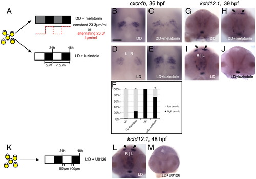
Melatonin signaling contributes to the timely differentiation of habenular neurons in constant darkness. (A) Embryos were either placed in DD and treated with melatonin or placed in LD and treated with the melatonin receptor antagonist luzindole. (B–C) Melatonin treatment under DD conditions phenocopies LD embryos: no excess precursors and (G–H) timely appearance of kctd12.1-positive neurons. (D–E) Conversely, antagonism of melatonin receptors under LD conditions results in a phenotype similar to DD embryos: accumulation of excess cxcr4b-positive precursors and (I–J) delayed appearance of kctd12.1-positive neurons. (F) Quantification of data represented in panels B–E. (K) Embryos in LD conditions were treated with the ERK1/2 phosphorylation inhibitor U0126. (L–M) A delay in the appearance of kctd12.1 neurons, similar to that seen in DD conditions, is observed. All views are frontal except A–D (dorsal views). Scale bar = 50 μm. |
Reprinted from Developmental Biology, 358(1), de Borsetti, N.H., Dean, B.J., Bain, E.J., Clanton, J.A., Taylor, R.W., and Gamse, J.T., Light and melatonin schedule neuronal differentiation in the habenular nuclei, 251-61, Copyright (2011) with permission from Elsevier. Full text @ Dev. Biol.