
Fig. 5
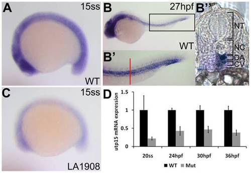
Analysis of endogenous utp15 expression by whole-mount in situ hybridization. A, utp15 is expressed ubiquitously in developing embryos at early stages. B, Expression was restricted to neural and vascular tissues after one day of development. B′, 2x magnification of the rectangular region indicated in (B), showing expression in the dorsal aorta and cardinal vein. Red bar, approximate location of the vibratome cross-section shown in (B′′). C–D, utp15 was dramatically reduced in mutant embryos, as shown by in situ hybridization (C) and qRT-PCR (D) analyses. A–C, Embryos shown are representative of triplicate experiments performed on a minimum of 5 embryos per condition. D, Data shown are the average fold changes in expression±SEM from two replicate qRT-PCR assays using pooled RNA from 20 embryos per condition. |
| Gene: | |
|---|---|
| Fish: | |
| Anatomical Terms: | |
| Stage Range: | 14-19 somites to Prim-25 |