|
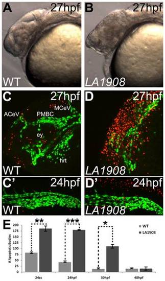
Apoptosis is pervasive in neural tissues of LA1908 mutant embryos. A–B, Brightfield phase-contrast imaging of mutant embryos revealed craniofacial disorganization and extensive cell death (hazy dark brown appearance in midbrain in B versus A). C–D′, Apoptosis is significantly induced in LA1908 mutant embryos (D, D′) relative to stage-matched WT siblings (C, C′). Green fluorescence marks the endothelial cells. Red fluorescence indicates apoptotic cells detected by TUNEL assay. ACeV, Anterior cerebral vein. ey, eye primordium. hrt, embryonic heart. MCeV, Middle cerebral vein. PMBC, Primordial midbrain channel. E, Quantitative and statistical analyses of apoptotic bodies at several stages of development in WT versus mutant embryos. * = p-value<0.05, ** = p-value<0.005, *** = p-value<0.00005 by two-tailed t-test, n = 3 embryos for each condition, data are average±SEM.
|