
Fig. 1
- ID
- ZDB-FIG-110816-31
- Publication
- Klaassen et al., 2011 - Synaptic transmission from horizontal cells to cones is impaired by loss of connexin hemichannels
- Other Figures
- All Figure Page
- Back to All Figure Page
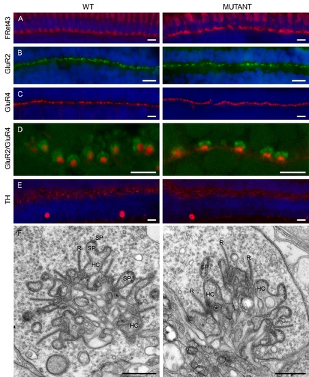
Localization of cellular markers in retinas from wild-type and mutant zebrafish. The organization of the outer retina was evaluated by the distribution of a number of cellular markers for double cones (A, FRet43) in red, horizontal cells (B, GluR2) in green, OFF-bipolar cells (C, GluR4) in red, and interplexiform cells (E, TH) in red. The blue stain is the nuclear marker DAPI. No difference in the distribution of these markers could be observed. In both preparations, GluR2 forms horseshoe-like structures in the OPL (B, D). Previous studies have shown that these structures are horizontal cell dendrites invaginating the cone terminal [57], [57], [58]. (E) TH labels interplexiform cells and the synapses these cells make onto horizontal cells. The immunoreactivity pattern is similar in wild-type and mutant retinas. These results indicate that all major neuron types in the OPL have developed normally. (A to E): Scale bar = 10 μm. (F) Ultrastructure of the cone synaptic terminal in wild-type (left) and mutant (right) retinas. No differences were found in the ultrastructural organization of the cone synaptic terminal. In both animal models, the characteristic shape of the synaptic triad was easily identified. R, synaptic ribbon; Sp, spinules; HC, horizontal cell; * bipolar cell. Scale bar = 0.5 μm. |
| Gene: | |
|---|---|
| Antibodies: | |
| Fish: | |
| Anatomical Terms: | |
| Stage: | Adult |