
Fig. 7
- ID
- ZDB-FIG-110712-26
- Publication
- Bruses, 2011 - N-cadherin regulates primary motor axon growth and branching during zebrafish embryonic development
- Other Figures
- All Figure Page
- Back to All Figure Page
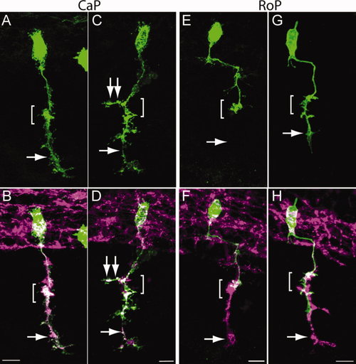
CaP and RoP motor axons grow abnormal branches in N-cadherin mutant embryos. Wild-type and cdh2hi3644Tg embryos were injected at the one-cell stage with a mix of two plasmids expressing Gal4 under the mnx1 promoter and pren-EGFP under a 14X-UAS element, fixed at 24 hpf, immunolabeled with SV2 and znp1 antibodies, and observed via confocal microscopy. A,C,E,G show pren-EGFP labeling; B,D,F,H show pren-EGFP (green) merged with SV2 and znp1 antibody labeling. A,B: Wild-type CaP motor neuron shows the characteristic morphology, with an axon extending from the spinal cord along the common pathway and into the myotome (arrow) ventral to the choice point (bracket). C,D: CaP motor neuron from a cdh2hi3644Tg embryo. The bracket indicates the choice point; the single arrow points to the axon in the ventral myotome; the double arrows point to an aberrant branch extending in the rostrocaudal axis. E,F: Wild-type RoP motor axon shows the characteristic caudal migration before turning ventrally into the myotome. The bracket indicates the choice point where the RoP axon normally stalled. The SV2 and znp1 labeling ventral to the choice point corresponds to the CaP motor axon. G,H: RoP motor neuron from a cdh2hi3644Tg mutant embryo. The bracket indicates the choice point, and the arrow points to aberrant growth of the axon into the ventral myotome together with the CaP motor axon. Rostral is to the left and dorsal is to the top. Scale bars = 10 μm in B (applies to A,B); 10 μm in D (applies to C,D); 10 μm in F (applies to E,F); 10 µm in H (applies to G,H). [Color figure can be viewed in the online issue, which is available at wileyonlinelibrary.com.] |