
Fig. 3
- ID
- ZDB-FIG-110706-32
- Publication
- Trikić et al., 2011 - Regulation of Zebrafish Hatching by Tetraspanin cd63
- Other Figures
- All Figure Page
- Back to All Figure Page
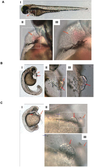
Morphology of cd63 knockdown. DIC microscopy images of the hatching glands of dechorinated 52 hour post fertilisation embryos. In each panel (A–C) I is x5 objective, white box indicates frame of II. II is x20 objective, white box indicates frame of III. III is x40 objective. II and III are montages of more than one focal plane. Red arrows point to the hatching gland, blue arrows indicate mislocalised hatching gland cells. A: Hatching gland typical of a LWT embryo. I is a montage of more than one focal plane. B: Moderately disrupted hatching gland typical after injection of 10 pg of MO4. C: Severely disrupted hatching gland typical after injection of 12 pg of MO4. |
| Fish: | |
|---|---|
| Knockdown Reagent: | |
| Observed In: | |
| Stage: | Long-pec |