
Fig. 5
- ID
- ZDB-FIG-110608-29
- Publication
- Knopf et al., 2011 - Bone Regenerates via Dedifferentiation of Osteoblasts in the Zebrafish Fin
- Other Figures
- All Figure Page
- Back to All Figure Page
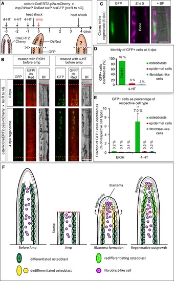
Genetic Fate Mapping of Osteoblasts in the Regenerating Fin (A) Experimental setup for genetic fate mapping using the osterix-driven inducible CreERT2 line (osterix:CreERT2-p2a-mCherry) in combination with the ubiquitous heat-shock inducible red to green responder line (hs:R to nG). (B) Stump osteoblasts contribute to cells in the 4 dpa regenerate. Whole-mount views of fin rays of ethanol (EtOH)-treated and 4-HT-treated fish, photographed after heat shock at 0 hpa and 4 dpa. Note that no recombined cells are found in EtOH-treated fins. Scale bar, 100 μm. (C) GFP+ cells in the regenerate visualized by anti-GFP immunostaining, which detects nuclear and some cytoplasmic GFP expressed from the hs:R to nG transgene, colabel with Zns 5 at 4 dpa. Scale bar, 50 μm. (D) The majority of GFP+ cells in the regenerate are Zns 5+ osteoblasts. Only few cells are found in the epidermis or are fibroblast-like cells. Quantification of sections of 4-HT-treated fins costained with Zns 5 (n = 5 fish, 10 rays, 61 sections with 1155 recombined cells). (E) Distribution of GFP+ cells in osterix:CreERT2-p2a-mCherry; hs:R to nG double-transgenic fins at 4 dpa. Of all cells in the regenerate, 0.4% are GFP+ in EtOH-treated fish versus 7.4% in 4-HT-treated fins. The fraction of GFP+ epidermal cells and fibroblast-like cells does not significantly differ between EtOH and 4-HT-treated fins, whereas 35-fold more GFP+ osteoblasts are found in 4-HT fins. Student′s t test, **p = 0.005 (nEtOH = 5 fish, 10 rays, 55 sections with 79 GFP+ cells; n4-HT = 5 fish, 10 rays, 61 sections with 1155 GFP+ cells). (F) Model of zebrafish fin bone regeneration based on our findings. Within 24 hr after amputation, distally located stump osteoblasts dedifferentiate and start to proliferate. Dedifferentiated osteoblasts migrate distally and form the lateral parts of the blastema. Thus, the blastema is derived from at least two cell types: osteoblasts and intraray fibroblast-like cells. Osteoblast-derived blastema cells do not transdifferentiate but differentiate only into osteoblasts. Thus, bone is regenerated from differentiated, mature osteoblasts. |
Reprinted from Developmental Cell, 20(5), Knopf, F., Hammond, C., Chekuru, A., Kurth, T., Hans, S., Weber, C.W., Mahatma, G., Fisher, S., Brand, M., Schulte-Merker, S., and Weidinger, G., Bone Regenerates via Dedifferentiation of Osteoblasts in the Zebrafish Fin, 713-724, Copyright (2011) with permission from Elsevier. Full text @ Dev. Cell