
Fig. 2
- ID
- ZDB-FIG-110407-23
- Publication
- Wühr M. et al., 2010 - A Model for Cleavage Plane Determination in Early Amphibian and Fish Embryos
- Other Figures
- All Figure Page
- Back to All Figure Page
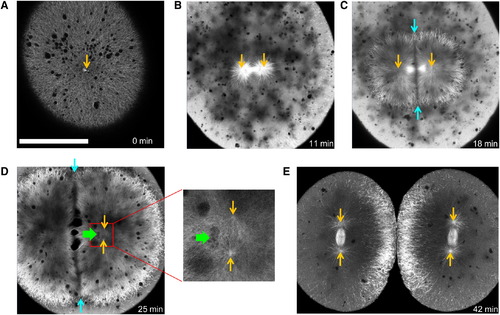
EMTB-3GFP Transgenic Zebrafish Embryos Allow Live Imaging of Microtubule Organization in Large Cells Orange arrows indicate positions of centrosomes.(A) Shortly after fertilization, sperm aster expands throughout the cell. The scale bar represents 200 μm. (B) Before metaphase, sperm aster breaks down and first mitotic spindle forms. (C) During anaphase-telophase, astral microtubules grow out, and centrosomes move apart. An interaction zone forms in the plane where sister asters contact each other (between blue arrows). (D) Centrosomes separate and align in the direction of the future spindle during late telophase (see enlargement). The centrosomes in the left aster are out of focus. Nuclei (green arrow) follow centrosomes, lagging behind. (E) Second mitotic spindles assemble after cytokinesis (E is taken from different embryo). See also Figure S2 and Movies S1 and S2. |