
Fig. 4
- ID
- ZDB-FIG-110112-18
- Publication
- Lin et al., 2010 - Loss of Cofilin 1 Disturbs Actin Dynamics, Adhesion between Enveloping and Deep Cell Layers and Cell Movements during Gastrulation in Zebrafish
- Other Figures
- All Figure Page
- Back to All Figure Page
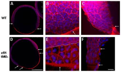
Knockdown of cfl1 enhances the filamentous actin (F-actin) formation and attenuates adhesion between the EVL and the DEL. Embryos at the 65%~70% epiboly stage were fixed, stained with rhodamine phalloidin and DAPI to reveal F-actin and nuclei, respectively, under confocal microscopy. Confocal images of whole-embryo sections at a lower magnification are shown for untreated (A) and cfl1 tMO1-injected embryos (B). In the untreated embryo (A), an arrow indicates synchronous leading edges of EVL and DEL whereas distinctly separated leading edges are obvious as pointed by arrows in cfl1 tMO1-injected embryos (B). At a higher magnification (B, C, E, F), F-actin was more condensed at the boundaries of blastomeres and the margin of blastoderm (arrows) of cfl1 morphants (E) compared to untreated embryos (B). The progression of the EVL margin (arrow) was more advanced than that of DEL (dashed line) in cfl1 morphants (E) compared to that of untreated embryos (B). From the side view (C, F), the EVL margin (upper arrow) was clearly advanced 2 cells ahead of DEL (lower arrow) in cfl1 morphants (F) that was not seen in untreated embryos (C). Scale bars: 25 μm for A and D; 250 μm for B, C, E, F. |
| Fish: | |
|---|---|
| Knockdown Reagent: | |
| Observed In: | |
| Stage: | Shield |