
Fig. 7
- ID
- ZDB-FIG-101208-43
- Publication
- Schneider et al., 2010 - Zebrafish Nkd1 promotes Dvl degradation and is required for left-right patterning
- Other Figures
- All Figure Page
- Back to All Figure Page
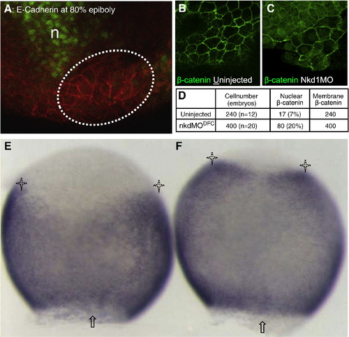
Nkd1 knockdown promotes nuclear β-catenin accumulation in DFCs and activates β-catenin transcriptional targets. Immunohistochemistry on embryos at 80% epiboly. (A) The DFC region (dashed lines) was identified by the position relative to the gsc-GFP positive region (green) and yolk, cell membrane is noted by E-cadherin distribution. (B) In wt DFC, β-catenin is detected mostly in plasma membrane. (C) In Nkd1MODFC embryos, β-catenin is detected at the plasma membrane and in the nuclei of cells in DFC cluster. (D) Summary of β-catenin-positive nuclei present in DFCs of wt and Nkd1MODFC embryos. n = notochord. *P < 6.8 × 10- 6, Fisher′s exact test. WMISH of axin2, dorsal side shown. (E) axin2 expression is excluded from the DFC region (arrow) with distinct lateral domains (asterisks) in ControlMODFC embryos. (F) Expanded axin2 expression is detected in Nkd1MODFC embryos. 91% show expanded domains, n = 33 compared to 7% in control MO, n = 27 (p-value = 2.1 × 10- 11). |
| Gene: | |
|---|---|
| Fish: | |
| Knockdown Reagent: | |
| Anatomical Term: | |
| Stage: | 90%-epiboly |
Reprinted from Developmental Biology, 348(1), Schneider, I., Schneider, P.N., Derry, S.W., Lin, S., Barton, L.J., Westfall, T., and Slusarski, D.C., Zebrafish Nkd1 promotes Dvl degradation and is required for left-right patterning, 22-33, Copyright (2010) with permission from Elsevier. Full text @ Dev. Biol.