|
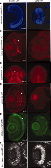
Defects in retinal cell differentiation in FucT8 morphant embryos. A:DAPI staining of eye cross-sections from 5-day-old control and FucT8 MO-injected embryos. Morphant embryos have a smaller retina, and although cell layers seem to be present, they are not as distinct as in control embryos. B,C: zn-5 immunostaining at 48 hpf identifies the RGCs (B, arrowhead) and optic chiasma (C, arrow) in control embryos, which are absent in morphant sections. One hundred percent of 20 injected embryos showed this phenotype. D: At 4 days, anti-CA-II antibody shows a lack of Müller glia (arrowhead) in the morphant retina. Eighty-five percent of 40 injected embryos showed this phenotype. E: zpr-1-positive photoreceptor cells are also severely reduced in FucT8 morphant eyes. Similar to 85% of 40 injected embryos showed this phenotype. F: BrdU incorporation illustrates increased cell proliferation in 28-hpf FucT8 morphant eyes. Eighty-two percent of 22 injected embryos showed this phenotype.
|