
Fig. 3
- ID
- ZDB-FIG-101021-33
- Publication
- Lyons et al., 2010 - Carboxypeptidase A6 in zebrafish development and implications for VIth cranial nerve pathfinding
- Other Figures
- All Figure Page
- Back to All Figure Page
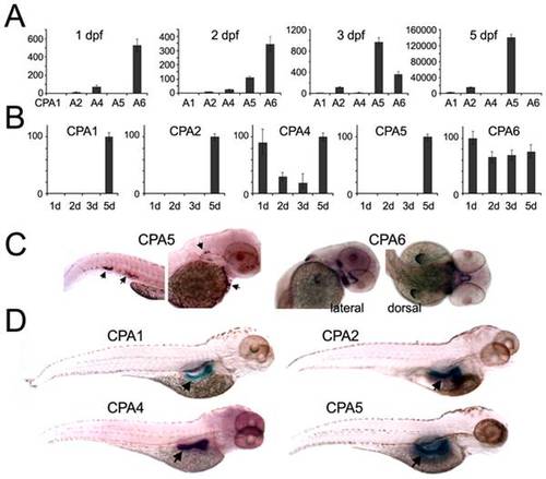
Temporal and spatial expression of zebrafish CPA genes. (A, B) qPCR was performed using cDNA prepared from zebrafish at the indicated developmental time points. All values were normalized to β-actin, and are shown in (A) as expression relative to CPA1 mRNA at 2 dpf, or in (B) as expression of each gene relative to its highest level (100%). (C, D) In situ hybridization was used to determine the spatial distribution of CPA mRNA expression. (C) At 2 dpf CPA5 mRNA (arrows) was detected in a mast cell population and CPA6 mRNA was seen in precursor tissues found in the stomodeum, posterior to the eyes, and in the pectoral fin buds. (D) At 4 dpf, mRNA for CPA1, 2, 4, and 5 were expressed in the pancreas. |
| Genes: | |
|---|---|
| Fish: | |
| Anatomical Terms: | |
| Stage Range: | Prim-5 to Day 5 |