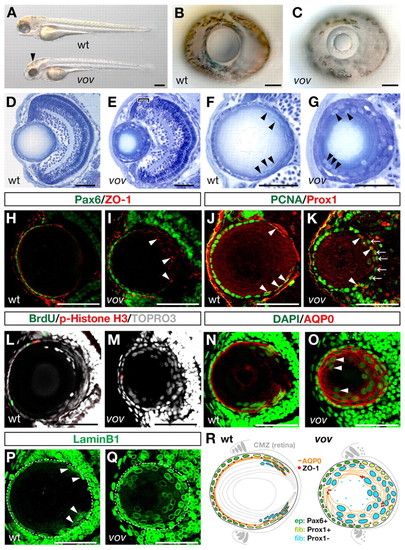
Zebrafish vov mutant shows defects in lens fiber cell differentiation. (A) Wild-type (wt) and vov mutant 72 hpf embryos. The vov mutant embryos exhibit hemorrhaging on the roof of the mid-hindbrain (arrowhead). (B,C) Wild-type (B) and vov mutant (C) eyes at 72 hpf. (D,E) Sections of 72 hpf wild-type (D) and vov mutant (E) eyes. Bracket indicates apoptosis near the retinal ciliary marginal zone (CMZ) (E). (F,G) Wild-type (F) and vov mutant (G) lens. Nuclei are flat and positioned along the posterior edge of the wild-type lens sphere (arrowheads, F), whereas nuclei are swollen and located in the anterior region surrounding a small lens fiber core in the vov mutant (arrowheads, G). (H,I) Expression of Pax6 (green) and ZO-1 (red) in 72 hpf wild-type (H) and vov mutant (I) lens. Arrowheads indicate aggregated ZO-1 signals in the posterior lens region of the vov mutant. (J,K) Expression of Pcna (green) and Prox1 (red) in 72 hpf wild-type (J) and vov mutant (K) lens. Arrowheads indicate the Pcna and Prox1 double-positive cells. Arrows indicate Pcna-positive cells located in the posterior edge of the lens sphere (K), most of which weakly express Prox1 (see Fig. S3D-D4 in the supplementary material). (L,M) Double labeling of 72 hpf wild-type (L) and vov mutant (M) lens with BrdU (green) and anti-pH3 antibody (red). Nuclei are stained with TOPRO3 (gray). (N,O) Expression of AQP0 (red) in 72 hpf wild-type (N) and vov mutant (O) lens. Nuclei are stained with DAPI (green). Arrowheads indicate nuclei of AQP0-positive lens fiber cells in the vov mutant, which are located in the anterior lens region (O). (P,Q) Expression of Lamin B1 in 72 hpf wild-type (P) and vov mutant (Q) lens. Arrowheads indicate wild-type lens fiber cell nuclei. White dashed lines indicate the outline of the lens sphere. (R) Schematic of wild-type and vov mutant lens. Lens epithelium (ep) expresses Pax6 (green) and its marginal cells are associated with retinal CMZ. After passing over the equator, lens epithelial cells transiently express Prox1 (light green) and differentiate into lens fiber cells (fib). Newly differentiating lens fiber cells (light blue) express AQP0 (orange) and their nuclei become flat and are subsequently denucleated. In the vov mutant, cell proliferation of lens epithelium is decreased and lens fiber cells are disorganized in morphology. ZO-1 signals are indicated by red circles. Anterior is left and dorsal is up in all the panels showing the lens (except in Fig. 3E,F and Fig. 6). Scale bars: 50 µm, except 250 µm in A.
|
