
Fig. 4
- ID
- ZDB-FIG-100719-23
- Publication
- Say et al., 2010 - A Functional Requirement for PAK1 Binding to the KH(2) Domain of the Fragile X Protein-Related FXR1
- Other Figures
- All Figure Page
- Back to All Figure Page
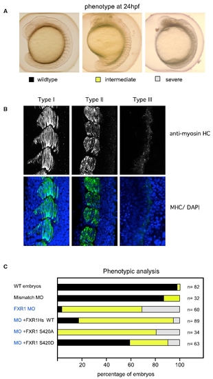
Rescue of FXR1 Depletion in Zebrafish Using Human FXR1 mRNA (A) Zebrafish embryos injected with synthetic morpholino-oligonucleotides (MO) against Zf FXR1; 2.5 ng was the lowest concentration that gave a high penetrance of phenotype (>90%). The organization of somites observed by bright field microscopy were scored as normal (black) when the chevron-shaped structures were clearly visible; intermediate (yellow) when the somites were clearly segmented but the shape abnormal; or severely disrupted (gray) when somite boundaries were poorly defined. (B) FXR1 leads to disorganization of muscle fibers. Muscle organization in the three classes of embryos illustrated in (A) was assessed by myosin heavy-chain (HC) staining and confocal imaging (5 μM section), with nuclei counterstained by DAPI. (C) Rescue of FXR1 loss by co-injection at the one-cell stage. Analysis of the phenotypes observed in embryos injected with FXR1 MO and co-injection with mRNA (100 ng) encoding wild-type FXR1, FXR1(S420A), or phospho-mimetic FXR1(S420D). The color coding for the bars indicates the embryos illustrated in (A). |
| Antibody: | |
|---|---|
| Fish: | |
| Condition: | |
| Knockdown Reagent: | |
| Anatomical Term: | |
| Stage: | Prim-5 |
| Fish: | |
|---|---|
| Knockdown Reagent: | |
| Observed In: | |
| Stage: | Prim-5 |