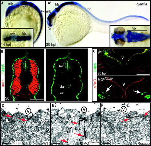
Loss of Claudin5a disrupts the neuroepithelial paracellular barrier function. (A and A′) Whole-mount in situ hybridization of claudin5a (cldn5a) expression at two developmental time points. Dorsal views onto the brain are shown magnified within the insets. (A) Before brain ventricle formation (14 hpf), cldn5a is strongly expressed within the developing central nervous system including the hindbrain (hb) and spinal cord (sc). Weaker expression of cldn5a is found within the dorsal midbrain region (mb). (A′) During ventricle expansion (30 hpf), cldn5a is strongly expressed within the spinal cord and the neuroepithelial ventricular zones of ventral hindbrain and midbrain. There is also strong expression in the forebrain (fb) ventricular zone. (B) Confocal microscopic images of cross-sections through the trunk region. Strong expression of Cldn5 proteins is present within the spinal cord (sc) and the dorsal aorta (da) but not the cardinal vein (cv) at 30 hpf. (C) Injection of MOcldn5a efficiently blocks expression of Cldn5a protein within the apical neuroepithelium lining the brain ventricles (V). Endothelial expression of Cldn5b is not affected in cldn5a morphants (arrows). (D–F) Electron micrographs of neuroepithelial cells covering the cerebral ventricles (V) after intraventricular injection of the electron-dense molecule lanthanum nitrate. In the WT, paracellular clefts are tight for the tracer, which accumulates in a dot-like pattern at the TJ (arrows). Knock-down of cldn5a results in diffusion of lanthanum nitrate into the paracellular space between cells (arrows). In rescue embryos, in which the cldn5a knock-down was rescued by concomitant cldn5a mRNA injection, electron dense material is confined to apico-lateral membranes of neuroepithelial cells similar to the distribution in WT embryos. (Scale bars: B and C, 50 μm; D, 2 μm.)
|
