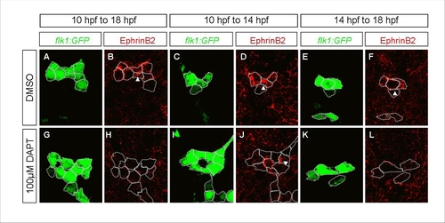
FIGURE

Fig. S5

ID
ZDB-FIG-091215-60
Publication
Lee et al., 2009 - Notch Signaling Functions as a Cell-Fate Switch between the Endothelial and Hematopoietic Lineages
Other Figures
All Figure Page
Back to All Figure Page
Fig. S5

A Distinct Role for Notch Signaling in the Formation of Endothelial Progenitors
Transverse sections of embryos treated with DMSO (A-F) or DAPT (G-L), visualized for kdrl:GFP ( and K) (green) and EphrinB2 (B, D, F, H, J and L) (red). Embryos were treated from 10 to 18 hpf (A-B, and G-H), from 10 to 14 hpf (C-D, and I-K), or from 14 to 18 hpf (E-F, and K-L). White dotted lines highlight the cell boundaries, and white arrowheads point to the expression of EphrinB2 within endothelial cells. DAPT treatment from 10 to 14 hpf leads to an increase in endothelial precursors, but does not affect EphrinB2 expression. Treatment from 14 to 18 hpf leads to a loss of EphrinB2 expression, but does not affect endothelial cell number.

Expression Data

Expression Detail
Antibody Labeling
Phenotype Data

Phenotype Detail
Acknowledgments
This image is the copyrighted work of the attributed author or publisher, and ZFIN has permission only to display this image to its users. Additional permissions should be obtained from the applicable author or publisher of the image. Full text @ Curr. Biol.