
Fig. 4
- ID
- ZDB-FIG-090504-46
- Publication
- Glasgow et al., 1997 - Neuronal and neuroendocrine expression of lim3, a LIM class homeobox gene, is altered in mutant zebrafish with axial signaling defects
- Other Figures
- All Figure Page
- Back to All Figure Page
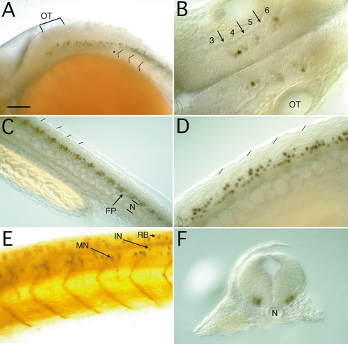
Expression of Lim3 in the hindbrain and spinal cord in 28-h embryos. Anterior is to the left except in F. (A) Lateral view of the hindbrain and anterior spinal cord. Dorsal is up. Lim3-positive cells in the hindbrain extend to rhombomere 4, just anterior to the otic vesicle, OT (brackets). Two rows of Lim3-positive cells are seen in the posterior hindbrain, one row appearing to be continuous with the ventral spinal cord cells. The first somite is marked with an asterisk, and the first three somite borders are indicated. (B) Ventral view of the hindbrain. Three clusters of Lim3-positive cells are seen in the center of rhombomeres 4, 5, and 6. Rhombomeres 3–6 are numbered, with arrows indicating the location of the rhombomere borders. OT, otic vesicle. (C) Lateral view of the spinal cord. Lim3-positive cells are arranged segmentally along the ventral spinal cord. The plane of focus is at a slight angle so that superficial structures are anterior and deeper structures are posterior. At the anteriormost side, somite boundaries are clearly seen (hash marks). Next, the Lim3-positive cells come into focus slightly above the midline in the ventral spinal cord. At the midline, the floor plate (FP) and notochord (N) are in focus. Toward the posterior, the Lim3-positive cells on the deeper side of the midline come into focus. (D) Closeup lateral view of the spinal cord. The stained nuclei of the segmentally arranged cells are clearly seen. (E) Lateral view of the spinal cord, double labeled with anti-Lim3 (purple) and zn12 (brown) antibodies. The zn12 monoclonal antibody recognizes the L2/HNK1 epitope on many neurons. Cells with Lim3-stained nuclei also have ventrally projecting axons that stain with zn12, whereas not all cells with zn12-labeled ventrally projecting axons express Lim3. An example of a double-labeled ventrally projecting motoneuron (MN) is indicated, along with a probable interneuron (IN) that is negative for Lim3 and positive for zn12. The soma and axons of the dorsally located Rohon–Beard (RB) sensory neurons label heavily with zn12. The zn12 immunoreactive myosepta are prominent. (F) Cross section through the anterior spinal cord. The Lim3-positive cells are in a ventrolateral position on both sides of the midline well separated from the floor plate cells and the notochord (N). Scale bar is 100 μm in A and C, 50 μm in B, D, and E, and 60 μm in F. |
| Gene: | |
|---|---|
| Antibodies: | |
| Fish: | |
| Anatomical Terms: | |
| Stage: | Prim-5 |
Reprinted from Developmental Biology, 192, Glasgow, E., Karavanov, A.A., and Dawid, I.B., Neuronal and neuroendocrine expression of lim3, a LIM class homeobox gene, is altered in mutant zebrafish with axial signaling defects, 405-419, Copyright (1997) with permission from Elsevier. Full text @ Dev. Biol.