
Fig. S3
- ID
- ZDB-FIG-090220-20
- Publication
- Ota et al., 2009 - The roles of the FGF signal in zebrafish embryos analyzed using constitutive activation and dominant-negative suppression of different FGF receptors
- Other Figures
- All Figure Page
- Back to All Figure Page
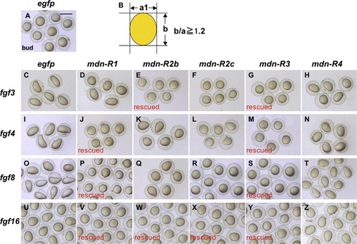
Embryos co-injected with mRNA for mdn-R and fgf genes. Zebrafish embryos were co-injected with mRNA (300–400 pg/embryo) for egfp or specified mdn-R genes together with different fgf mRNA at minimal amounts sufficient to attain complete dorsalization. All the fgf genes caused dorsalization, as revealed by elongation along the animal-vegetal axis at the bud stage (compare C, I, O, U with A), which was suppressed selectively by some of the mdn-R genes. The criterion of rescue was whether the ratio of the animal-vegetal axis (b) to the other axis (a) was reduced below 1.2 (B). ‘Rescue’ shows nearly complete rescue of embryos by mdn-R overexpression. |
Reprinted from Mechanisms of Development, 126(1-2), Ota, S., Tonou-Fujimori, N., and Yamasu, K., The roles of the FGF signal in zebrafish embryos analyzed using constitutive activation and dominant-negative suppression of different FGF receptors, 1-17, Copyright (2009) with permission from Elsevier. Full text @ Mech. Dev.