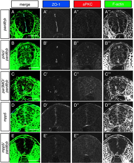
Role of the Par3/Par6/aPKC complex in apical membrane formation in the neural tube. All images represent transverse sections between the first and sixth somite stained for ZO-1 (blue), aPKC (red), and filamentous actin (green) at 24 hpf. (A) pard6γbs441 mutant neural tubes show a reduced amount of apical membranes. (B) pard6γbs441/prkci double mutants show a more severe reduction in apical membrane formation than pard6γbs441 mutants. (C) pard6γbs441 mutants injected with a pard3 morpholino show a more severe reduction in apical membrane formation than pard6γbs441 mutants. (D) mpp5 mutant neural tubes do not show an obvious severe reduction in apical membrane formation. (E) pard6γbs441/mpp5 double mutants show a more severe reduction in apical membrane formation than pard6γbs441 or mpp5 single mutants. In addition, pard6γbs441/prkci and pard6γbs441/mpp5 double mutants as well as pard6γbs441 mutants injected with a pard3 morpholino exhibit a severe epithelial disorganization in the neural tube. Scale bars represent 50 μm.
|
