
Fig. 4
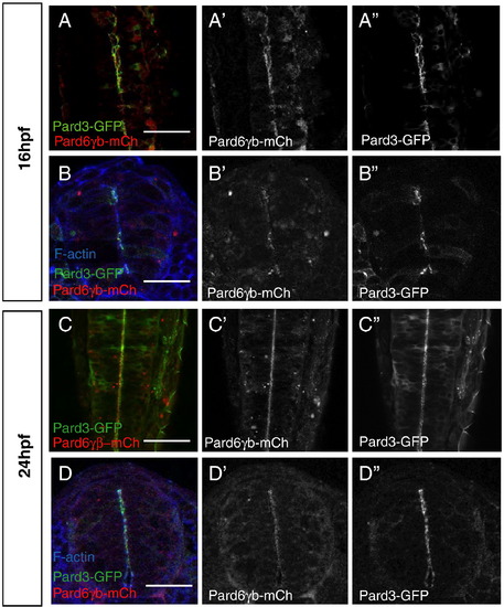
Distribution of Pard6γb during neurulation. (A–D) All embryos were injected with pard3-gfp and pard6γb-mch mRNA. (A, C) Optical sections through the neural tube of live embryos (dorsal views between the first and sixth somite, anterior up). (B, D) Transverse sections through the neural tube of embryos between the first and sixth somite stained for filamentous actin. (A, B) At 16 hpf, Pard6γb-mCh begins to localize with Pard3-GFP to the forming apical membranes of the neural tube. (C, D) At 24 hpf, Pard6γb-mCh localizes with Pard3-GFP along the maturing apical membranes of the neural tube. Scale bars represent 50 μm. |
Reprinted from Developmental Biology, 324(1), Munson, C., Huisken, J., Bit-Avragim, N., Kuo, T., Dong, P.D., Ober, E.A., Verkade, H., Abdelilah-Seyfried, S., and Stainier, D.Y., Regulation of neurocoel morphogenesis by Pard6gammab, 41-54, Copyright (2008) with permission from Elsevier. Full text @ Dev. Biol.