
FIGURE
Fig. 9
- ID
- ZDB-FIG-080916-26
- Publication
- Park et al., 2000 - Analysis of upstream elements in the HuC promoter leads to the establishment of transgenic zebrafish with fluorescent neurons
- Other Figures
- All Figure Page
- Back to All Figure Page
Fig. 9
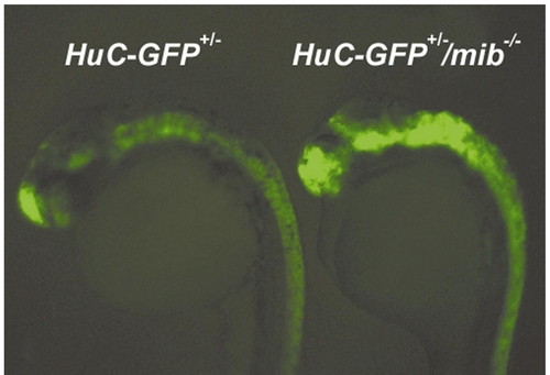
Characterization of living mib mutant transgenic embryos by GFP fluorescence. The neurogenic phenotype in 2-day-old HuC-GFP+/-/mib-/- zebrafish embryo seen by GFP fluorescence with a Leica MZFLIII fluorescence stereomicroscope (right), compared with a heterozygotic wild-type HuC-GFP+/- transgenic embryo (left). |
Expression Data
Expression Detail
Antibody Labeling
Phenotype Data
Phenotype Detail
Acknowledgments
This image is the copyrighted work of the attributed author or publisher, and
ZFIN has permission only to display this image to its users.
Additional permissions should be obtained from the applicable author or publisher of the image.
Reprinted from Developmental Biology, 227(2), Park, H.-C., Kim, C.-H., Bae, Y.-K., Yee, S.-Y., Kim, S.-H., Hong, S.-K., Shin, J., Yoo, K.-W., Hibi, M., Hirano, T., Miki, N., Chitnis, A.B., and Huh, T.-L., Analysis of upstream elements in the HuC promoter leads to the establishment of transgenic zebrafish with fluorescent neurons, 279-293, Copyright (2000) with permission from Elsevier. Full text @ Dev. Biol.