
FIGURE
Fig. 6
- ID
- ZDB-FIG-080916-22
- Publication
- Park et al., 2000 - Analysis of upstream elements in the HuC promoter leads to the establishment of transgenic zebrafish with fluorescent neurons
- Other Figures
- All Figure Page
- Back to All Figure Page
Fig. 6
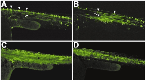
In vivo functional analysis of HuC promoter constructs in living zebrafish. Promoter deletion constructs for ΔHind (A), ΔBst (B), ΔSac (C, D) were microinjected to the one-cell (A, B, C) or four-cell embryos (D). Fluorescence images of GFP in 48 hpf living zebrafish embryos were captured by the laser confocal microscopy. GFP expression in the neuronal cells and muscle cells are indicated by the arrowheads and arrows, respectively. |
Expression Data
Expression Detail
Antibody Labeling
Phenotype Data
Phenotype Detail
Acknowledgments
This image is the copyrighted work of the attributed author or publisher, and
ZFIN has permission only to display this image to its users.
Additional permissions should be obtained from the applicable author or publisher of the image.
Reprinted from Developmental Biology, 227(2), Park, H.-C., Kim, C.-H., Bae, Y.-K., Yee, S.-Y., Kim, S.-H., Hong, S.-K., Shin, J., Yoo, K.-W., Hibi, M., Hirano, T., Miki, N., Chitnis, A.B., and Huh, T.-L., Analysis of upstream elements in the HuC promoter leads to the establishment of transgenic zebrafish with fluorescent neurons, 279-293, Copyright (2000) with permission from Elsevier. Full text @ Dev. Biol.