
Fig. 1
- ID
- ZDB-FIG-080909-21
- Publication
- Trede et al., 2008 - Zebrafish mutants with disrupted early T-cell and thymus development identified in early pressure screen
- Other Figures
- All Figure Page
- Back to All Figure Page
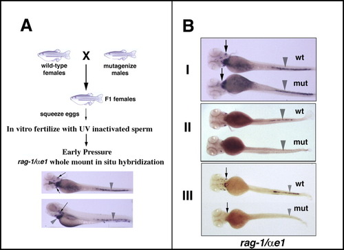
Design of screen and classes of mutants obtained. A:Schematic diagram of screen design. For details, refer to the Results and Experimental Procedure sections. Whole-mount in situ hybridization (WISH) at 5 days postfertilization (dpf) wild-type (wt) larvae with rag-1 and αe1 probes shows expression of rag-1 in bilateral thymi (arrows) and αe1 in the heart and tail (arrowhead). B:Three classes of mutants were observed in the screen. In each panel the wild-type (wt) larvae are on top, the mutant (mut) larvae are on the bottom. I. Lymphoid mutants showed normal expression of αe1 (arrowheads) but absence of rag-1 expression (arrows) compared with wt controls (top panel). II. Erythroid mutants showed normal expression of rag-1, but defective expression of αe1 (middle panel). III. Mutants showing defects in both rag-1 and αe1 expression point to a possible defect in the early hematopoietic compartment (bottom panel). |