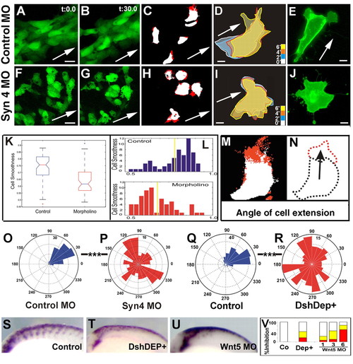
Syn4 and PCP signaling control the orientation of cell protrusions. (A,B,F,G) Time-lapse analysis of the mandibular NC stream of a sox10:egfp embryo at 18 hpf for 30 minutes. The initial and final frames are shown. Arrow indicates the expected direction of migration based on the orientation of the embryo. Cell extensions are shown in C and H in red. (D,I) Outline of individual cells at different times (minutes). (E,J) NC cells cultured in vitro, expressing membrane GFP. (A-E) Control MO; (F-J) syn4 MO. Scale bars: 25 μm in A,F; 10 μm in D,E,I,J. (K,L) Quantification of cell smoothness (CS) of NC cells. (K) Box-plot showing the distribution of CS values for control and syn4 MO-injected cells. A significant difference was observed (P<0.005). (L) Histogram of CS for control and syn4-MO injected cells. (M,N) Analysis of CE. (M) Two consecutives frames were subtracted, in a manner that the new growing area is shown in red and the unchanged area in white. A vector between the centroid of the cell and the centroid of the red area was drawn (arrow in N). (O-R) Rose plots showing the orientation of CE. (O) Control MO; (P) syn4 MO; (Q) control embryo; (R) embryo injected with DshDep+. Note the difference in scale. Scale bar: 20 μm; ***P<0.005. (S-V) Non-canonical Wnt signaling is required for zebrafish neural crest migration. Lateral views of dorsal neural tubes of 20-somite zebrafish embryos are shown after crestin staining by in situ hybridization. Dorsal to the top; anterior to the left. (S) Control; (T) 200 pg of DshDep+ mRNA; (U) 3 ng of wnt5 MO. (V) Percentage of inhibition in trunk NC migration. White, no inhibition; yellow, mild inhibition; red, strong inhibition.
|
