FIGURE

Fig. 1

ID
ZDB-FIG-080430-10
Publication
Ozbudak et al., 2008 - Notch Signalling Synchronizes the Zebrafish Segmentation Clock but Is Not Needed To Create Somite Boundaries
Other Figures
All Figure Page
Back to All Figure Page
Fig. 1

DAPT Treatment Blocks Notch Signalling Rapidly
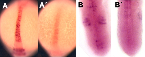
DAPT causes loss of her4 expression (A) in the neural tube and (B) in the anterior PSM (where her4 is normally expressed in a faint but detectable stripe). Wild-type embryos were treated with 100 μm DAPT or with DMSO (control) medium for 1 h and stained by ISH for her4. In each panel, the DMSO control is shown on the left, the DAPT-treated embryo on the right. Treatment was begun at the 13-somite stage for (A) and at the 15-somite stage for (B). For each case, at least 14 embryos were examined, and typical specimens were selected for this illustration.

Expression Data
Gene:
Fish:
Condition:
Anatomical Terms:
Stage: 14-19 somites

Expression Detail
Antibody Labeling
Phenotype Data

Phenotype Detail
Acknowledgments
This image is the copyrighted work of the attributed author or publisher, and ZFIN has permission only to display this image to its users. Additional permissions should be obtained from the applicable author or publisher of the image. Full text @ PLoS Genet.