
Fig. 4
- ID
- ZDB-FIG-080425-4
- Publication
- Lieschke et al., 2002 - Zebrafish SPI-1 (PU.1) marks a site of myeloid development independent of primitive erythropoiesis: implications for axial patterning
- Other Figures
- All Figure Page
- Back to All Figure Page
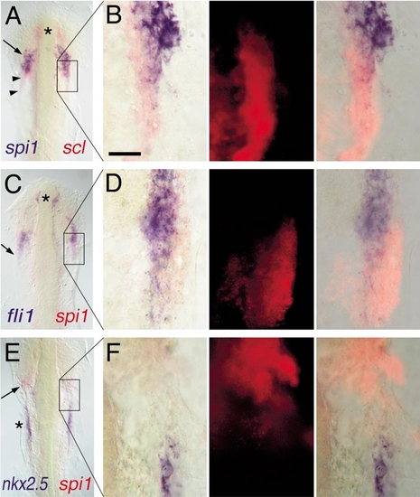
Expression of spi1 relative to other markers of lateral plate mesoderm fates. Two-color in situ hybridization gene expression analysis of wild-type zebrafish embryos (12 somite, flat mount, anterior up). Each panel in the left column shows a low-power view of the head and rostral lateral plate (A, C, E), and the corresponding three panels to the right show high-magnification views (63x water immersion lens) of particular regions of interest in which the expression of scl or spi1 has been detected by fluorescence of Fast Red using an RITC filter set (B, D, F). In places, the presence of high levels of blue NBT/BCIP precipitate quenches the fluorescent signal. (A, B) spi1 (blue) and scl (red). Asterisk marks rostral portion of scl+/spi1- cells, arrow shows domain of scl+/spi1+ cells, and arrowheads show scl+/spi1- cells caudal to the domain of coexpressing cells. (C, D) fli1 (blue) and spi1 (red). Asterisk marks rostral fli1+/spi1- cells, and the arrow shows that, although fli1 and spi1 are largely coexpressed, at the lateral margin, some fli1-/spi1+ cells are evident. (E, F) nkx2.5 (blue) and spi1 (red). The heart anlage marked by nkx2.5 (asterisk) is separate and caudal to the region of spi1 expression (arrow). Scale bar in (B), 30 μm. |
Reprinted from Developmental Biology, 246(2), Lieschke, G.J., Oates, A.C., Paw, B.H., Thompson, M.A., Hall, N.E., Ward, A.C., Ho, R.K., Zon, L.I., and Layton, J.E., Zebrafish SPI-1 (PU.1) marks a site of myeloid development independent of primitive erythropoiesis: implications for axial patterning, 274-295, Copyright (2002) with permission from Elsevier. Full text @ Dev. Biol.