
Fig. 3
- ID
- ZDB-FIG-080425-3
- Publication
- Lieschke et al., 2002 - Zebrafish SPI-1 (PU.1) marks a site of myeloid development independent of primitive erythropoiesis: implications for axial patterning
- Other Figures
- All Figure Page
- Back to All Figure Page
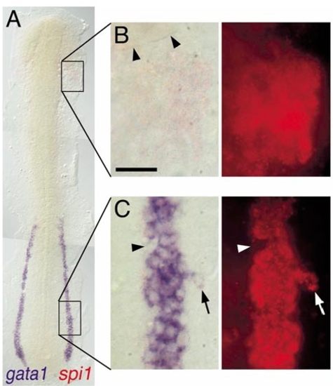
Expression of spi1 relative to gata1 in the lateral plate mesoderm. Two-color in situ hybridization gene expression analysis of wild-type zebrafish embryo (12 somite, flat mount, rostral up). (A)Alow-power view spanning the entire lateral plate mesoderm (LPM). (B, C) High-magnification views (63x water immersion lens) of regions boxed in (A), in which the expression of spi1 has been detected by fluorescence of Fast Red using a RITC filter set. (B) spi1+gata1- cells in the rostral LPM. Arrowheads indicate the caudal margin of the eye. (C) spi1+gata1+ cells in the caudal LPM. Arrowhead indicates an indentation to the otherwise uniform LPMgata1+ domain, due to several cells that express neither gata1 nor spi1. Arrow indicates a lateral irregularity in the LPM gata1 domain, due to several cells that express both gata1 and spi1. Scale bar in (B), 30 μm. |
Reprinted from Developmental Biology, 246(2), Lieschke, G.J., Oates, A.C., Paw, B.H., Thompson, M.A., Hall, N.E., Ward, A.C., Ho, R.K., Zon, L.I., and Layton, J.E., Zebrafish SPI-1 (PU.1) marks a site of myeloid development independent of primitive erythropoiesis: implications for axial patterning, 274-295, Copyright (2002) with permission from Elsevier. Full text @ Dev. Biol.