|
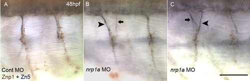
Aberrant exit points of secondary motoneuron (SMN) axons in Nrp1a-knockdown embryos. (A) Axons of primary motoneurons (PMNs) and SMNs at 48 hpf in a control MO-injected embryo. PMN axons were labeled with a brown stain, recognized by mAb Znp1, but not by Zn5 (Zeller et al., 2002). SMN axons were stained in blue-black, recognized by Zn5 (Fashena and Westerfield, 1999), regardless of Znp1 reactivity. SMN axons extended ventrally along with PMN axons using an exit point common to PMN and SMN. (B) Double exit phenotype of SMN axons was observed in Nrp1a-knockdown embryos at 48 hpf. Generally, one axonal bundle of SMNs was thicker (arrowhead) than the others (arrow), regardless of its anterior or posterior position. (C) Another example of a double exit phenotype of SMN. Scale bar: 50 μm.
|