
FIGURE
Fig. 1
Fig. 1
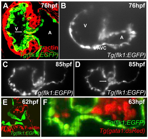
Zebrafish valves form through invagination. Larvae were stained with rhodamine-phalloidin and imaged using confocal microscopy at 76 (A) or 62 (E) hpf, or imaged using SPIM at 76 (B), 85 (C,D) or 63 (F) hpf. Although by confocal microscopy a superior endocardial cushion appears to be present (panel A), SPIM imaging shows that this structure is actually a valve leaflet (B-D). Arrow in panel A points to the presumed endocardial cushion and that in D to the valve leaflet. No intermediate stage of mesenchymal cells was observed by either confocal (E) or SPIM imaging (F). A, atrium; V, ventricle. |
Expression Data
Expression Detail
Antibody Labeling
Phenotype Data
Phenotype Detail
Acknowledgments
This image is the copyrighted work of the attributed author or publisher, and
ZFIN has permission only to display this image to its users.
Additional permissions should be obtained from the applicable author or publisher of the image.
Full text @ Development