|
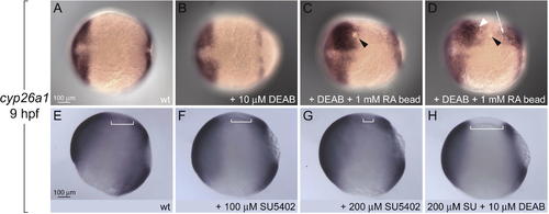
Regulation of cyp26a1 by RA and Fgfs (A and B) Anterior and marginal expression domains of cyp26a1 expression are unchanged by 10 μM DEAB. Anterior is to the left in all panels. (C and D) RA beads (black arrowheads) induce cyp26a1 expression in DEAB-treated embryos far from (white arrowhead and arrow in [D]), but not adjacent to the bead. (E–H) Fgf signaling sets the posterior boundary of cyp26a1 expression. (E) cyp26a1 expression in wild type (wt) (lateral view, anterior to the left). (F and G) Expression in SU5402-treated embryos shifts posteriorly in a concentration-dependent manner (compare brackets in [E], [F] and [G]). (H) The posterior shift caused by blocking Fgf signaling is abolished by DEAB treatment.
|