|
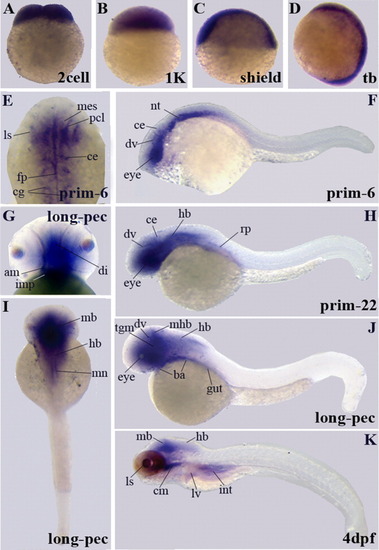
A-K: Gene expression patterns of enigma homologue variants encoding the LIM domain. Similar to the PDZ domain encoding variant, the results of whole-mount in situ hybridization analysis is shown for the eight stages indicated. F,H,J,K:Embryos at prim-6, prim-22, long-pec, and 4 dpf stages are shown in lateral view. I: A dorsal view of the long-pec stage is shown. E,G: Higher magnifications of expression domains in the head are presented for prim-6 in dorsal view (E) and for the long-pec stage in frontal view (G). am, adductus mandibulae; ba, branchial arches; cm, cephalic musculature; ce, cerebellum; cg, cranial ganglia; di, diencephalon; dv, diencephalic vein; fp, floor plate; hb, hindbrain; imp, intermandibularis posterior; int, intestine; mes, mesencephalon; mb, midbrain; mhb, midbrain-hindbrain boundary; ls, lens; lv, liver; mn, motor neurons; nt, notochord; pcl, proliferative cell layer; rf, roof plate; tct, tectum; tgm, tegmentum.
|