
Fig. 8
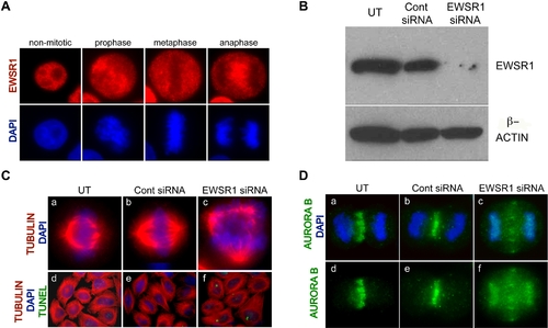
EWSR1 siRNA transfected Hela cells Display Mitotic Defects. A. EWSR1 localization in Hela cells during non-mitotic and mitotic-stages. EWSR1 was visualized by anti-EWSR1 antibody (red) and DNA was stained with DAPI (blue). B. EWSR1 protein (top panel) and β-Actin (bottom panel) visualized by western blotting. C. Representative images of mitotic spindles from (a and d) UT, (b and e) Cont siRNA and (c and f) EWSR1 siRNA transfected Hela cells. DNA was stained with DAPI (blue), the spindles were visualized by α-Tubulin (red) and the apoptotic cells were visualized by TUNEL assay (green). D. Representative images of Aurora B (green) with DNA (blue) (a–c), and Aurora B (green) without DNA (d–f). (a and d) UT, (b and e) Cont siRNA and (c and f) EWSR1 siRNA transfected Hela cells. UT: untransfected, Cont siRNA: control siRNA transfected, EWSR1 siRNA: EWSR1 siRNA transfected Hela cells. |