
FIGURE
Fig. 3
- ID
- ZDB-FIG-071006-2
- Publication
- Arnaout et al., 2007 - Zebrafish model for human long QT syndrome
- Other Figures
- All Figure Page
- Back to All Figure Page
Fig. 3
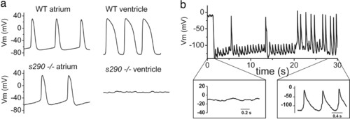
APs recorded from explanted embryonic zebrafish hearts. (a) (Upper) Representative spontaneous APs recorded from wild-type (WT) atrium (Left) and ventricle (Right) at 48 hpf. (Lower) Spontaneous APs recorded from kcnh2s290 mutant atrium (Left). Recordings of transmembrane voltage (V m) from mutant ventricle (Right) revealed marked membrane depolarization and the absence of action potentials. (b) V m recorded from kcnh2s290 mutant ventricle. Intracellular injection of hyperpolarizing current (-100 pA) caused membrane hyperpolarization and allowed for the generation of spontaneous APs. |
Expression Data
Expression Detail
Antibody Labeling
Phenotype Data
| Fish: | |
|---|---|
| Observed In: | |
| Stage: | Long-pec |
Phenotype Detail
Acknowledgments
This image is the copyrighted work of the attributed author or publisher, and
ZFIN has permission only to display this image to its users.
Additional permissions should be obtained from the applicable author or publisher of the image.
Full text @ Proc. Natl. Acad. Sci. USA