
Fig. 1
- ID
- ZDB-FIG-070806-1
- Publication
- Arnaout et al., 2007 - Zebrafish model for human long QT syndrome
- Other Figures
- All Figure Page
- Back to All Figure Page
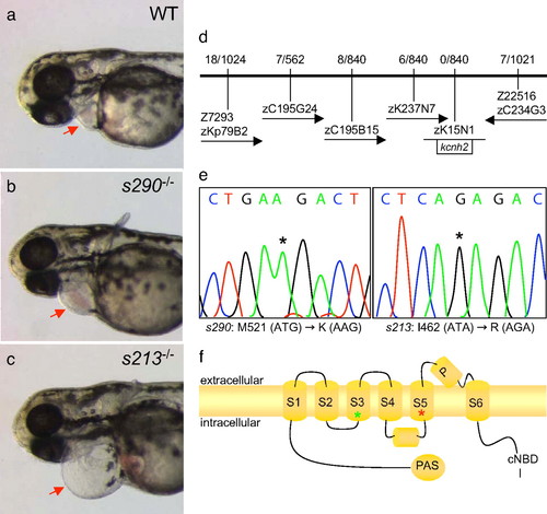
Molecular analysis of two kcnh2 mutant alleles. (a–c) Wild-type and mutant embryos at 48 hpf, lateral oblique views, anterior to the left. Compared with wild type (WT) (a), s213 (b), and s290 (c), mutants exhibit a silent ventricle and, therefore, absent cardiac output and pronounced pericardial edema (red arrows). (d) The mutations map to linkage group 3. Flanking CA repeat markers and markers from selected BACs are shown, with the number of recombinants for the given number of meioses. The s213 and s290 mutations map to the potassium channel gene kcnh2. (e) Sequence analysis of kcnh2 in s213 mutants reveals a T–G transition at codon 462, resulting in an Ile–Arg substitution. For s290, a T–A transversion at codon 521 results in a Met–Lys substitution. (f) Schematic diagram showing the modular structure of Kcnh2. A Kcnh2 subunit consists of six transmembrane domains: the S1–S4 domains sense membrane potential, whereas the S5–S6 domains form the K-selective pore. Green and red stars represent the s213 and s290 mutation sites, respectively. |
| Fish: | |
|---|---|
| Observed In: | |
| Stage: | Long-pec |