
Fig. 1
- ID
- ZDB-FIG-070917-105
- Publication
- Connors et al., 2006 - Temporal and spatial action of Tolloid (Mini fin) and Chordin to pattern tail tissues
- Other Figures
- All Figure Page
- Back to All Figure Page
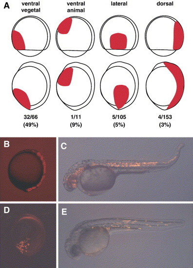
Tolloid activity is required in ventral-vegetal cells. (A) Schematic of cell transplantations (location of wild type cells in red) in gastrula or bud stage embryos, N values and percent of rescue observed. The dorsal group also includes dorsolateral clones (0/32 rescued). Not included are 7/30 (23%) cases of rescue seen in transplants that were scored as ventrolateral at the margin. (B) Fluorescence image of an embryo at bud stage with cells located dorsally and (C) the same embryo at 24 hpf displaying the mfn phenotype, indicating no rescue. (D) Fluorescence image of an embryo at bud stage with transplanted cells located ventrally, and (E) the same embryo at 24 hpf shows rescue of the mfn phenotype. (C, E) Fluorescence images are overlaid on the bright field image to show the location of transplanted cells at 24 hpf. All are lateral views except (D), which is slightly ventral-lateral. (A, B, D) Dorsal is to the right and anterior is up. (C, E) Anterior to the left. |
Reprinted from Developmental Biology, 293(1), Connors, S.A., Tucker, J.A., and Mullins, M.C., Temporal and spatial action of Tolloid (Mini fin) and Chordin to pattern tail tissues, 191-202, Copyright (2006) with permission from Elsevier. Full text @ Dev. Biol.