
Fig. 3
- ID
- ZDB-FIG-070815-15
- Publication
- Schibler et al., 2007 - A screen for genetic defects of the zebrafish ear
- Other Figures
- All Figure Page
- Back to All Figure Page
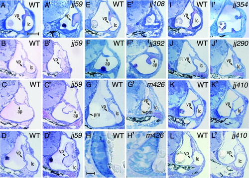
Histological analysis of morphology mutants. Transverse plastic section were prepared through ears of larval zebrafish. The first, the third and the fifth column from the left display sections of wild-type animals, while the second, the fourth, and the sixth column display sections of their mutant siblings at corresponding stages. The hako mimijj59 mutant phenotype is characterized by a thickening of inner ear pillars at 72 hpf (A′ compare to the wild type in A). By 120 hpf (B′), the ventral pillar of the kmijj59 ear frequently expands, blocking the lumen of the lateral canal (compare to the wild type in B). Panel (C′) displays a malformed kmijj59 anterior pillar characterized by an abnormal outgrowth at 120 hpf (compare to C). Another example of the kmijj59 mutant, featuring a bulb-like outgrowth instead of the ventral pillar is shown in (D′). The ear of mikre uchojj108 is strongly reduced in size at 72 hpf and its walls are thickened (E′, compare to E). Also at 72 hpf, smojj392 mutants (F′) feature a malformation of anterior pillar shape and size. At 144 hpf, antm426 mutant embryos (G′), display a reduced otic vesicle that is almost completely filled by the otolith. Enlargements of wild-type and antm426 mutant posterior maculae are shown in (H) and (H′), respectively. Note the malformation of the posterior macula in the antm426 mutant. Panel (I′) displays the pata nopojj354 mutant ear. At 72 hpf, its ventral pillar is incompletely formed. The ear of frostbitejj290 mutant is reduced at 144 hpf (J′, compare to J). In, ale uchojj410, epithelial protrusions that form the ventral pillar are thickened and incompletely formed at 72 hpf. (K′). By 120 hpf, however, ale uchojj410 ventral pillar displays a largely normal shape (L′). The following structures of the ear are indicated with arrows: ap, anterior pillar; vp, ventral pillar; pm, posterior macula. “lc” indicates lateral canal. In all panels, dorsal is up and lateral is right. Scale bar in (H) equals 10 μm, and applies to panels (H) and (H′). Scale bar in (A) equals 50 μm, and applies to all remaining panels. |
| Fish: | |
|---|---|
| Observed In: | |
| Stage Range: | Protruding-mouth to Day 6 |
Reprinted from Mechanisms of Development, 124(7-8), Schibler, A., and Malicki, J., A screen for genetic defects of the zebrafish ear, 592-604, Copyright (2007) with permission from Elsevier. Full text @ Mech. Dev.