
Fig. 3
- ID
- ZDB-FIG-070421-2
- Publication
- Shankaran et al., 2007 - Completing the set of h/E(spl) cyclic genes in zebrafish: her12 and her15 reveal novel modes of expression and contribute to the segmentation clock
- Other Figures
- All Figure Page
- Back to All Figure Page
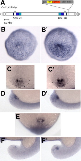
Expression of the MMLV-retroviral enhancer trap CLGY-521 during somitogenesis. (A) Schematic representation of the CLGY-521 insertion site 535 bp 5′ to the her15b gene on Chr 11. LTR = long terminal repeat, gata2P = zebrafish gata2 promoter, YFP = yellow fluorescent protein. (B, B′) Representative embryos from weak and strong expression classes, respectively, showing yfp mRNA distribution at bud stage; vegetal views, dorsal up. (C, C′) Weak and strong expression classes at 12 somites, dorsal view of tail bud. (D, D′) Same embryos in panels C, C″ viewed laterally, to highlight the ventral core of expression. (E) Embryo from panels C, D viewed axially from posterior, to reveal the ring-like shape of expression domain. (F, F′) Weak and strong expression classes at 17 somites, lateral view of tail bud. |
Reprinted from Developmental Biology, 304(2), Shankaran, S.S., Sieger, D., Schroter, C., Czepe, C., Pauly, M.C., Laplante, M.A., Becker, T.S., Oates, A.C., and Gajewski, M., Completing the set of h/E(spl) cyclic genes in zebrafish: her12 and her15 reveal novel modes of expression and contribute to the segmentation clock, 615-632, Copyright (2007) with permission from Elsevier. Full text @ Dev. Biol.