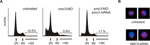
Cell DNA content analysis and chromosomal abnormalities following SMC3 knockdown. A) Cell cycle FACS analysis of zebrafish embryonic cells. Embryos at 1–2 cell stage were injected with 1 nl Danieau buffer (left-hand side) or 4 ng of smc3-MO (center) or 4 ng of smc3-MO together with 250 pg smc3 mRNA (right-hand side). Batches of 30 embryos at 24 hpf were incubated in 0.05% trypsin 15 min at 25°C and the cells dissociated by pipetting through a large bore glass pipette. After fixation in 70% ethanol at 4°C overnight, cells were stained with propidium iodine/RNase, and the DNA cell content analyzed by flow cytometry. Gated data were used for analysis. Note the presence of a population of aneuploid (DNA > 4) cells in embryos following Smc3 knockdown. B) Analysis of the centrosome organization in human cells. Human 293 cells were transfected with 50 ng/ml of SMC3-siRNA and after 24 h plated on a poly-lysine-coated coverslip. Fourty-eight h later the cells were fixed in 4% para-formaldehyde, permebilized and then incubated with 1:1000 anti-human γ-tubulin antibody. Following incubation with Alexafluor 567-conjugated anti-rabbit IgG (Molecular Probes) and DNA staining with DAPI, the cells were examined under a fluorescence/UV microscope and the red (Alexafluor 567) and blue (DAPI) signals combined.
|
