
Fig. 1
- ID
- ZDB-FIG-070418-39
- Publication
- Choi et al., 2007 - FoxH1 negatively modulates flk1 gene expression and vascular formation in zebrafish
- Other Figures
- All Figure Page
- Back to All Figure Page
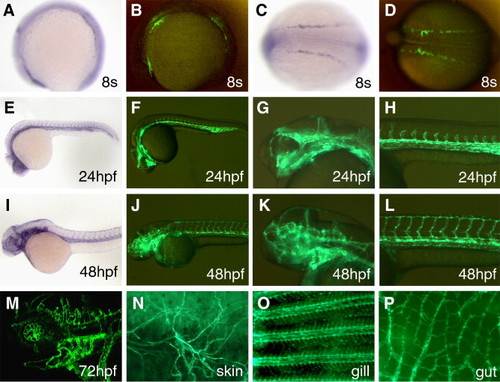
GFP expression in TG(flk1:GFP)la116 embryos. (A–D) GFP signals driven by the flk(6.4)-GFP transgene can be detected in TG(flk1:GFP)la116 embryos as early as the 8-somite stage. flk1 expression is detected in three patches of cells by in situ hybridization (A). A similar pattern is seen in TG(flk1:GFP)la116 embryos (B). Lateral views are shown in panels A and B and the dorsal views are shown in panels C and D. Anterior to the left. (E–H) GFP expression pattern of TG(flk1:GFP)la116 embryos at 24 hpf (F) resembles the flk1 pattern detected by in situ hybridization (E). Higher magnification images of the head and trunk are shown in panels G and H, respectively. (I–L) GFP expression pattern of TG(flk1:GFP)la116 embryos at 48 hpf (J) resembles the flk1 pattern detected by in situ hybridization (I). Higher magnification images of the head and trunk are shown in panels K and L, respectively. (M) Confocal image of GFP expression in endothelial cells in the brain and brachial arches in 3-day-old TG(flk1:GFP)la116 embryos. (N–P) GFP expression remains active in adult TG(flk1:GFP)la116 fish. Images show GFP signals in endothelial cells in the skin (N), gill (O) and gut (P). |
| Gene: | |
|---|---|
| Fish: | |
| Anatomical Terms: | |
| Stage Range: | 5-9 somites to Long-pec |
Reprinted from Developmental Biology, 304(2), Choi, J., Dong, L., Ahn, J., Dao, D., Hammerschmidt, M., and Chen, J.N., FoxH1 negatively modulates flk1 gene expression and vascular formation in zebrafish, 735-744, Copyright (2007) with permission from Elsevier. Full text @ Dev. Biol.