|
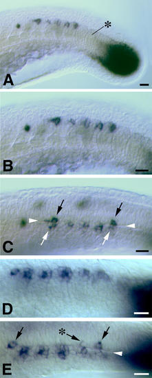
The juxtaposition of spt expression in newly formed somites and the developing CNS at approximately 22 hpf. In all images dorsal is uppermost and rostral is to the left. A, B and C are views from one embryo. A and B show the appearance from a lateral view of the tail in the region of the "somitic trail" of spt expression. A black asterisk indicates the most recently formed somite. spt expression is concentrated to the dorsocaudal extremity of somites. In an optical (DIC) section through the same embryo viewed from a dorsolateral perspective (C), the basal lamina separating the developing CNS and the somitic mesoderm can be seen clearly (arrowheads). Cells expressing spt in the developing CNS (black arrows) are juxtaposed to somitic cells expressing spt (white arrows). The "somitic trail" region of a second embryo is shown in D (lateral view) and E (dorsolateral view). The black asterisk in E indicates a neural cell expressing a lower level of spt. Scale bars equal 20 μm.
|