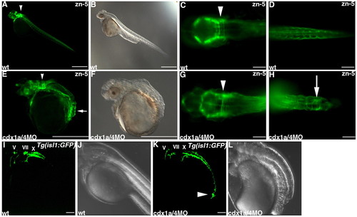
Ectopic formation of hindbrain neurons in embryos lacking Cdx1a and Cdx4. (A-H) Detection of hindbrain commissure neurons by immunostaining with monoclonal antibody zn-5 of wild-type control (A-D) and cdx1a/4 morphant embryos (E-H) at 3 days post fertilization (dpf). (B,E) Bright field images. Lateral views (A,B,E,F) and high-magnification dorsal views of hindbrain (C,G) and tail regions (D,H) with anterior to the left. zn-5-positive commissure neurons can be recognized by their axonal structures in the hindbrain regions (arrowheads) and ectopically in the posteriormost neural tissue (arrows). (I-L) Detection of cranial motoneurons in control Tg(isl1:GFP) embryos and Tg(isl1:GFP) embryos that received cdx1aMO and cdx4MO at 48 hpf. Bright field images (J,L). The position of trigeminal (V), facial (VII) and vagal (X) motor nuclei was indicated. A cluster of the GFP+ neurons with their axons were detected in the posteriormost region of cdx1a/4 morphant embryos (arrowhead, K). Scale bars: 500 μm in A,B,E,F; 100 μm in C,D,G,H,I,K.
|
