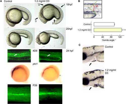
(A) Fertilized zebrafish eggs were dechorionated and placed in buffer containing 1.2 mg/ml sonicated vitamin D3, starting the continuous incubation between the 32- and 64-cell stages. At the different developmental stages (as indicated), the embryos were analysed, revealing persistent somite malformations, a ventrally curved body, poorly developed myoseptum, an aberrant extension of the yolk tube, a prominent dorsal midbrain, and a reduced trunk. The wild-type Engrailed staining (4D9) of muscle pioneers and surrounding cells in control-treated animals was not observed upon vitamin D3 treatment. ISH for ptc1 mRNA showed a strongly reduced signal in vitamin D3–treated animals compared with controls. Staining with the F59 antibody revealed that slow muscle fibres were disturbed in number and orientation in the treated animals. (B) At 18 hpf, embryos were photographed in detail and somite angles were determined (as shown in top panel). The measurement was taken of an individual somite between dorsal and ventral portions of the vertical myoseptum for corresponding somites between the wild-type and vitamin D3–treated embryos. Shown are the mean angles (±SEM; n = 3) of wild-type control- and vitamin D3–treated embryos. C) Detail of 4-dpf embryo. Note the slightly enlarged pericardial cavity in vitamin D3–treated animals. The orientation of all images is a lateral view, anterior to the left.
|
