
Fig. 6
- ID
- ZDB-FIG-060627-15
- Publication
- Chen et al., 2004 - Three modules of zebrafish Mind bomb work cooperatively to promote Delta ubiquitination and endocytosis
- Other Figures
- All Figure Page
- Back to All Figure Page
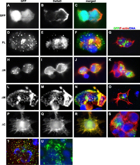
Mib-Delta interaction alters their subcellular distribution. The immunofluorescent images in each row show the subcellular localization of Mib or its deletion mutants in the presence or absence of DeltaD. The first column from the left shows the subcellular localization of Mib and its deletion mutants. The second column shows the subcellular localization of DeltaD. Third column shows the merged images of the first and second columns plus nuclear stain (blue). The rightmost column shows the merged images of Mib and its deletion mutants (green) in the absence of DeltaD. (A–C) Distribution of DeltaD and EGFP when co-expressed. (D–F) Co-localization of full-length Mib and DeltaD. (H–J) MibΔN and DeltaD do not co-localize. (L–N) MibΔM co-localizes with, but fails to enhance the internalization of DeltaD. (P–R) MibΔC co-localizes with DeltaD, and is distributed on the membrane and in perinuclear regions. (T) Distribution of full-length Mib partially overlaps that of Texas-Red-conjugated Dextran. (U) Full-length Mib, when expressed in zebrafish embryos after synthetic RNA injection, is also distributed in a punctuate manner. |
Reprinted from Developmental Biology, 267(2), Chen, W., and Casey Corliss, D., Three modules of zebrafish Mind bomb work cooperatively to promote Delta ubiquitination and endocytosis, 361-373, Copyright (2004) with permission from Elsevier. Full text @ Dev. Biol.