|
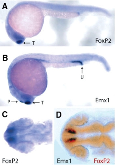
Expression of FoxP2 during zebrafish embryogenesis. (A) Lateral view of in situ hybridization of FoxP2 probe to a 20-somite zebrafish embryo. The expression is in the dorsal telencephalon (arrow). (B) Lateral view of in situ hybridization of Emx1 probe to a 20-somite zebrafish embryo. Expression is in the dorsal telencephalon (T), pineal gland (P) and the urogenital opening (U). (C) Dorsal view of the head region from a 20-somite zebrafish embryo hybridized with a FoxP2 probe. (D) Double in situ hybridization of a FoxP2 probe (red) and Emx1 (black) to the head region of a 20-somite zebrafish embryo. Dorsal view.
|