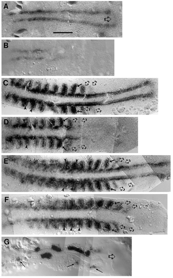
Dorsal views of the trunk regions showing MyoD expression during somite formation and development. 10.5 hour (A,B), 12.5 hour (C.D) and 14.5 hour (E.F,G) wild-type (A,C,E) ntl (B,D,F), or spt (G) embryos were dissected, yolk removed from under the trunk, and the tissue viewed and photographed under DIC optics. All embryos are shown with anterior to the left. An open arrow in A points to the region of expression connecting the two parallel rows of expressing cells. In C-F, open arrows indicate prospective somites either unsegmented or with only partially formed posterior furrows, solid arrows point to the most recently fully formed furrows, and arrowheads indicate older somites with changed shape and MyoD expression pattern. In G, arrows show expressing cells remote from the notochord, open arrow shows thick spade tail area devoid of expression of the gene. All panels are composites of views in which the plane of the notochord is kept in focus. Scale bar in A equals 100 µm, all panels at same magnification.
|
