
Fig. S2
- ID
- ZDB-FIG-050721-2
- Publication
- Rhinn et al., 2005 - Positioning of the midbrain-hindbrain boundary organizer through global posteriorization of the neuroectoderm mediated by Wnt8 signaling
- Other Figures
- All Figure Page
- Back to All Figure Page
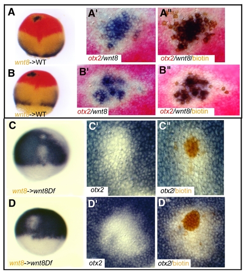
wnt8 expression does not self-induce and Wnt8 can repress otx2 in Df(Wnt8) host embryos. (A,B) Embryos containing cells derived from injected embryos with a lineage tracer (brown) and wnt8 RNA (400 pg) into the animal pole of a wild-type host embryo. (A′-B′′) Close-up of transplanted cells (A′,B′) before biotin staining and (A′′,B′′) after biotin staining. The embryos are stained for wnt8 (blue) and otx2 (red). No wnt8 expression is observed around the transplanted clone (brown) suggesting that the repression of otx2 is caused by by Wnt8 produced by the transplanted cells (n=15/15). (C,D) Embryos containing cells derived from injected embryos with a lineage tracer (brown) and wnt8 RNA (400 pg) into the animal pole of a host embryo mutant for wnt8 (Df(Wnt8)). (C′-D′′) Close up view of transplanted (C′,D′) before biotin staining and (C",D") after biotin staining. Wnt8 from the transplanted cells represses otx2 as no Wnt8 protein can be produced by the host embryo (mutant analysed n=10/10). |