
Fig. 5
- ID
- ZDB-FIG-050425-9
- Publication
- Down et al., 2005 - Cloning and expression of the large zebrafish protocadherin gene, Fat
- Other Figures
- All Figure Page
- Back to All Figure Page
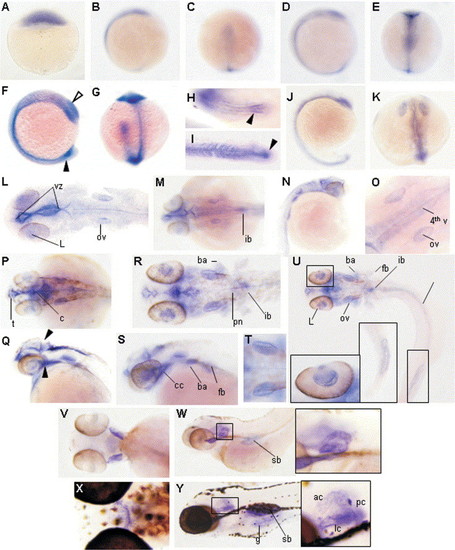
Expression of fat RNA by in situ hybridisation. A DIG labelled probe against EGFR5-FC2 of fat was used at a variety of developmental time points. (A) Expression was first seen at 4 hpf in the deep cells of the dome stage embryo, which may include the cells of the yolk syncytial layer. Expression is not then detected until after epiboly (B), (C) at 10 hpf where here is weak staining of the basal layers of the presumptive head, notochord and tail bud. With the onset of segmentation, expression is increased along the lengthening embryonic axis, and can clearly be seen in the furrow forming the notocord, head and tail structures (E). Expression is also becoming evident in the developing optic primordium, otic cup and tail bud, which is yet to extend away from the yolk (D). By 14 hpf we have strong expression throughout the embryo with the levels increasing in the optic cup, otic placode, extending tail bud, and may include Kupffer′s vesicle and the chordo-neural hinge (F), (G). The tail bud at this stage has prominent staining in the mesenchyme (arrowhead), with bilateral areas of medial somitic epithelium also staining (H). By 22 hpf only the most distal somites display expression on their posterior edge, compacting notocord mesenchyme is still strongly expressed (arrowhead) (I). At 17 hpf, staining is still present in the notochord, tail bud and head structures (J). By 22 hpf the optic primordium can be clearly seen and expression increases outlining the forming ventricles of the brain (K). (L) Represents an embryo at 26 hpf, in which the yolk has been removed and embryo flattened, a composite of 15 overlayed shots was used to capture the full depth of the embryo. Expression is now restricted to the lens of the eye (L), the otic vesicles (ov) and the lining of the ventricles from the telencephalon through to the cerebellum and 4th ventricle, encompassing the ventricular zone. Dorsally, 33 hpf expression is still prominent in the lining of the ventricles and ear and becomes apparent in the forming intestinal bulb (ib) (M). Laterally at this stage the forming fin bud is also noticeable and the notocord is still weakly positive (N). At high power it is possible to see that it is the lining of the 4th ventricle and the otic cups that are expressing (O). At 42 hpf expression persists in the lens, lining of the ventricles and otic cups, laterally the posterior tectum and the cranial cavity are outlined (arrowheads) cerebellum (c), telencephalon (t) (P), (Q). Expression at 50 hpf includes the posterior branchial arches (ba), fin bud (fb), intestinal bulb (ib), otic vesicle (ov), pronephros (p), spinal cord (sc), pronephric duct/gut (pd/g), cranial cavity (cc) and the ventricular lining. (R) Is a composite of 21 overlayed shots, inset displays staining specifically in the lining of the lens of the eye and the lining of the posterior widening gut (R), (U), and (S). Although at 50 hpf the sensory patches have formed it is the otic capsule epithelial lining that is ubiquitously expressing fat (T). By 100 hpf expression is lost in the ventricles and persists only in the brachial/gill arches swim bladder (sb) and most prominently in the sensory lining of the ear (inset) (V), (W). At 150 hpf expression is still evident in the gill arches and the sensory patches of the ear (inset) including the anterior (ac) and posterior crista (pc) and the lateral commissure (lc) but is also more evident in the lining of the swim bladder (sb) and gut (g) (X), (Y). |
Reprinted from Gene expression patterns : GEP, 5(4), Down, M., Power, M., Smith, S.I., Ralston, K., Spanevello, M., Burns, G.F., and Boyd, A.W., Cloning and expression of the large zebrafish protocadherin gene, Fat, 483-490, Copyright (2005) with permission from Elsevier. Full text @ Gene Expr. Patterns