
Fig. 3
- ID
- ZDB-FIG-050304-2
- Publication
- Iovine et al., 2005 - Mutations in connexin43 (GJA1) perturb bone growth in zebrafish fins
- Other Figures
- All Figure Page
- Back to All Figure Page
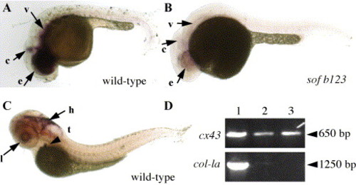
Expression of connexin 43 in embryos. In situ hybridization was completed using a cx43 probe against the cx43 3′ UTR. (A) Expression of cx43 in wild-type embryo at 24 hpf. e, eyes; c, cerebellum; v, vasculature. (B) Expression of cx43 in sof embryo at 24 hpf. e, eyes; c, cerebellum; v, vasculature. (C) Expression of cx43 in wild-type larvae at 72 hpf. Embryos were treated with PTU to prevent the production of melanin and facilitate the identification of stained structures. l, lens epithelium; h, hindbrain; t and arrowhead, thymus. (D) RT-PCR from 72 hpf whole embryos (lane 1), 72 hpf embryonic hearts (lane 2), and adult hearts (lane 3). The top panel shows amplification from cx43-3′ UTR primers, the bottom panel shows amplification from col-1a primers. Approximate sizes of amplified products are shown on the right (arrowhead). |
| Gene: | |
|---|---|
| Fish: | |
| Anatomical Terms: | |
| Stage Range: | Prim-5 to Adult |
Reprinted from Developmental Biology, 278(1), Iovine, M.K., Higgins, E.P., Hindes, A., Coblitz, B., and Johnson, S.L., Mutations in connexin43 (GJA1) perturb bone growth in zebrafish fins, 208-219, Copyright (2005) with permission from Elsevier. Full text @ Dev. Biol.