
- Title
-
Hdac11 inhibits apoptosis in zebrafish cells under cold stress via downregulating pdcd6 through its downstream enhancer
- Authors
- Ma, M., Luo, J., Li, X., Deng, R., Jiang, H., Han, B., Zhang, J.
- Source
- Full text @ Mol. Biol. Rep.

Hdac11 negatively regulates pdcd6 and apoptosis in zebrafish cells. (a, b) ZF4 cells were pre-treated with 1 µM JB3-22 or DMSO for 2 h, or transfected with pcDNA3.1(-) (Vector) or pcDNA3.1(-)-flag-hdac11-WT (OE-hdac11) for 8 h, then cultured at 28 °C–18 °C (cold stress) for 24 h. Apoptosis was detected with Annexin V-FITC/PI staining. (c) Relative mRNA level of hdac11 in ZF4 cells after transfection with pcDNA3.1(-)-flag-hdac11-WT for 24 h. (d) Relative mRNA level of pdcd6 in ZF4 cells after treatment with 1 µM JB3-22 for 24 h or transfected with pcDNA3.1(-)-flag-hdac11-WT (OE-hdac11) for 36 h. (e, f) Relative mRNA level of hadc11 and pdcd6 in WT and hdac11−/− zebrafish liver. Data are represented as mean ± SD of three independent experiments, and P values are calculated using Student’s t-test (n.s. P > 0.05; *P < 0.05; **P < 0.01; ***P < 0.001; ****P < 0.0001). |
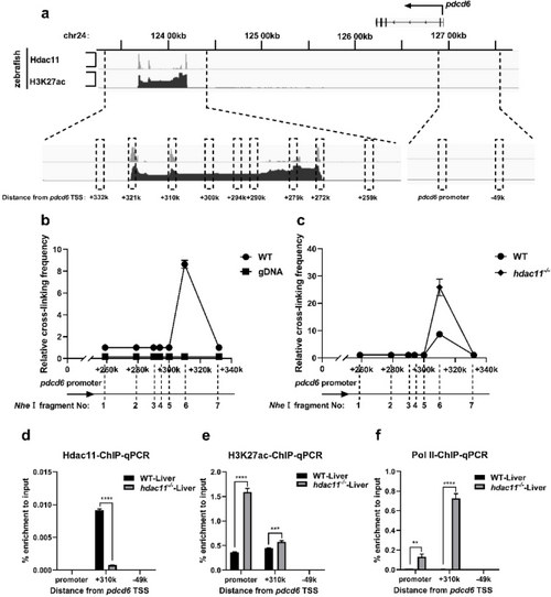
The knockout of hdac11 promotes interaction between pdcd6 + 310k region and pdcd6 promoter. (a) ChIP-seq data showed the enrichment of Hdac11 and H3K27ac at the pdcd6 locus. (b, c) 3 C-qPCR showed interaction frequency between indicated pdcd6 downstream regions and pdcd6 promoter in WT and hdac11−/− zebrafish liver. The Nhe Ⅰ fragment numbers represented different restriction sites, 1: +259k, 2: +279k, 3: +290k, 4: +294k, 5: +300k, 6: +310k, 7: +332k. (d, e, f) ChIP-qPCR showed the enrichment of Hdac11 (d), H3K27ac (e) and Pol Ⅱ (f) at indicated regions in zebrafish liver. Data are represented as mean ± SD of three independent experiments, and P values are calculated using Student’s t-test (**P < 0.01; ***P < 0.001; ****P < 0.0001) |
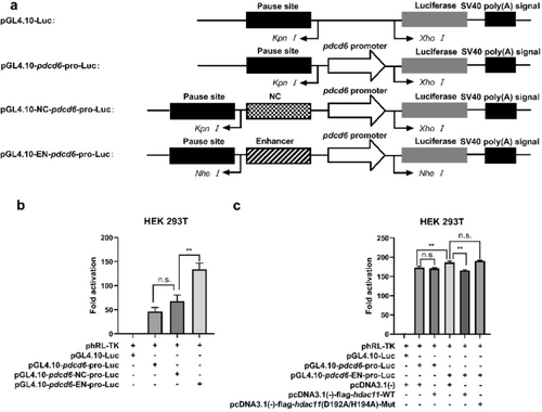
Hdac11 inhibits pdcd6 promoter activity through pdcd6 + 310k enhancer. (a) The structure diagram of indicated pGL4.10 plasmids. (b, c) HEK 293 T cells were transfected with indicated plasmids, then luciferase signal was detected. Data are represented as mean ± SD of three independent experiments, and P values were calculated using Student’s t-test (n.s. P > 0.05; **P < 0.01). |
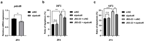
Inhibition of Hdac11 induces apoptosis through pdcd6 in ZF4 cells. (a) The mRNA level of pdcd6 was detected after ZF4 cells were transfected with 100 pmol sipdcd6 or siNC for 24 h. (b, c) ZF4 cells were pre-treated with 1 µM JB3-22 for 2 h, or transfected with 100 pmol sipdcd6, or siNC for 8 h, then cultured at 28 °C–18 °C (cold stress) for 24 h, respectively. Apoptosis was detected by Annexin V-FITC/PI staining. Data are represented as mean ± SD of three independent experiments, and P values were calculated using Student’s t-test (n.s. P > 0.05; **P < 0.01; ***P < 0.001; ****P < 0.0001). |